Ligand accessibility is an important parameter when evaluating the performance of magnetic beads used for antibody purification. Although a bead preparation may contain a high concentration of immobilized Protein A, not every ligand molecule is necessarily available to bind immunoglobulin G (IgG). Some binding sites may become sterically hindered, improperly oriented, or positioned within regions that antibodies cannot easily reach.
When you measure ligand accessibility, you gain a clearer understanding of how effectively your purification material captures antibodies. This information helps you distinguish between total ligand loading and functional binding capacity, identify potential manufacturing inconsistencies, and select suitable materials for reproducible purification workflows.
If you work with antibody capture applications, evaluating Magnetic Protein A Beads can help you assess whether the immobilized ligand provides sufficient accessible binding sites for your experimental requirements.
1. Understand Total Ligand Loading and Accessible Ligand Density
Before measuring ligand accessibility, distinguish between total Protein A loading and functional ligand availability.
Total ligand loading represents the amount of Protein A immobilized on the magnetic bead surface. Accessible ligand density describes the fraction of immobilized Protein A that remains available for interaction with its antibody target.
These measurements are not interchangeable. A bead preparation may contain substantial Protein A but demonstrate relatively low IgG-binding capacity because of steric hindrance, unfavorable ligand orientation, or restricted surface access.
To evaluate both properties, consider measuring:
- Total immobilized Protein A concentration.
- Functional IgG-binding capacity.
- Accessible ligand percentage.
- Antibody capture efficiency.
- Binding consistency across bead batches.
Comparing these parameters provides a more complete assessment of bead functionality.
2. Measure Functional Binding Capacity Using IgG
One practical approach to estimating ligand accessibility involves measuring the amount of IgG captured under controlled conditions.
Prepare a known concentration of purified IgG and incubate it with a measured quantity of magnetic beads. Maintain consistent buffer composition, pH, temperature, incubation time, and mixing conditions.
After incubation, separate the beads magnetically and measure the remaining IgG concentration in the supernatant using a suitable analytical method, such as UV absorbance at 280 nm or a validated protein assay.
Calculate the amount of captured IgG using:
\[ Q = \frac{(C_i-C_f)\times V}{m} \]
Where:
- \(Q\) = IgG-binding capacity (mg/g of beads).
- \(C_i\) = Initial IgG concentration (mg/mL).
- \(C_f\) = Final IgG concentration (mg/mL).
- \(V\) = Sample volume (mL).
- \(m\) = Magnetic bead mass (g).
This measurement estimates functional binding capacity rather than directly quantifying accessible Protein A molecules. However, it provides valuable information about how effectively the immobilized ligand participates in antibody capture.
3. Evaluate Ligand Accessibility Through ELISA
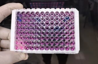
An ELISA-based assay can help determine whether immobilized Protein A retains accessible binding regions.
You can use an antibody that specifically recognizes an exposed Protein A epitope. Incubate the magnetic beads with the primary antibody, wash away unbound material, and introduce an enzyme-conjugated secondary antibody when required.
After adding the appropriate substrate, measure the resulting signal using a microplate reader.
A stronger signal generally indicates greater accessibility of the recognized Protein A epitope, provided that nonspecific binding and assay conditions are controlled.
This approach helps you investigate:
- Surface exposure of immobilized Protein A.
- Differences between bead preparation batches.
- Changes in ligand accessibility after storage.
- Potential steric interference caused by immobilization.
Remember that ELISA measures accessibility of the particular epitope recognized by your detection antibody. It does not necessarily represent the accessibility of every IgG-binding site.
4. Compare Ligand Accessibility at Different IgG Concentrations
A single IgG concentration may not reveal the complete binding behavior of your beads.
Perform an adsorption equilibrium experiment using multiple IgG concentrations while maintaining consistent bead quantities and incubation conditions.
Measure the remaining IgG in each supernatant and calculate the corresponding binding capacities. Plot the amount of captured IgG against the initial IgG concentration.
You can use a Langmuir adsorption model to estimate maximum binding capacity (Qmax) when the experimental data reasonably fit its assumptions.
This approach helps you identify whether increasing IgG concentrations produce additional binding or whether the available functional binding sites have approached saturation.
Research on magnetic Protein A purification has demonstrated the usefulness of adsorption equilibrium experiments and Langmuir modeling for characterizing antibody-binding capacity.
5. Examine Ligand Orientation and Steric Hindrance
Ligand orientation directly influences how effectively Protein A interacts with IgG.
If immobilization chemistry attaches Protein A in an unfavorable orientation, some antibody-binding regions may become difficult to access.
Similarly, excessive ligand density can create steric interference between neighboring Protein A molecules.
To investigate these effects, compare bead preparations produced using different immobilization conditions, coupling chemistries, or ligand concentrations.
Evaluate their functional IgG-binding capacities under identical experimental conditions.
You should also examine whether changes in immobilization conditions affect antibody recovery and binding consistency.
Comparative research on Protein A immobilization has shown that ligand orientation and surface density can influence IgG-binding performance.
6. Monitor Accessibility After Storage and Repeated Use
Ligand accessibility can change during storage, handling, and repeated purification cycles.
Protein A may experience structural changes, while bead aggregation or surface contamination can reduce effective contact between antibodies and binding sites.
To monitor these changes, perform functional binding assays at predefined intervals.
Compare fresh beads with stored or regenerated samples using identical IgG concentrations, incubation times, and analytical methods.
Record:
- Initial and remaining IgG concentrations.
- Functional binding capacity.
- Antibody recovery percentage.
- Nonspecific protein adsorption.
- Bead aggregation and magnetic separation performance.
These measurements help you identify whether reduced purification performance results from ligand deterioration, physical bead changes, or unfavorable experimental conditions.
7. Improve Experimental Consistency
Reliable ligand accessibility measurements require standardized experimental conditions.
Use the same bead mass, sample volume, buffer composition, mixing speed, and incubation temperature across experiments.
Include appropriate controls, such as beads without Protein A, assay blanks, and reference bead preparations.
Perform replicate measurements to evaluate reproducibility. When possible, use an independent method to verify important findings.
For example, compare total Protein A loading with IgG-binding capacity and an epitope-specific accessibility assay. These measurements provide complementary information rather than interchangeable estimates.
Conclusion
Measuring ligand accessibility helps you understand the functional performance of magnetic affinity purification materials beyond their total Protein A content. By combining IgG-binding capacity measurements, ELISA-based accessibility testing, adsorption equilibrium analysis, and storage stability assessments, you can evaluate how effectively immobilized Protein A supports antibody capture.
For dependable purification results, select Magnetic Protein A Beads based on functional binding performance, reproducibility, and compatibility with your experimental workflow.
Lytic Solutions, LLC provides molecular biology tools and affinity reagents for research and protein purification applications.
Contact us today to discuss your protein purification requirements.
Frequently Asked Questions
What is ligand accessibility in Magnetic Protein A Beads?
Ligand accessibility refers to the fraction of immobilized Protein A molecules whose binding regions are physically available to interact with IgG during antibody purification.
How do you measure Protein A ligand accessibility?
You can estimate functional accessibility through IgG-binding capacity assays, ELISA-based epitope detection, and comparative adsorption studies under controlled experimental conditions.
Is ligand density the same as ligand accessibility?
No. Ligand density measures the amount of Protein A present on the bead surface, whereas ligand accessibility describes how much of that immobilized Protein A remains functionally available for antibody binding.
How does ELISA help evaluate Protein A accessibility?
ELISA detects accessible Protein A epitopes using specific antibodies and enzyme-generated signals, helping you compare surface accessibility between different bead preparations.
Why does Protein A orientation matter during immobilization?
Unfavorable orientation can conceal antibody-binding regions or position them close to the bead surface, reducing the effective interaction between Protein A and IgG.
Can excessive Protein A loading reduce ligand accessibility?
Yes. Excessive ligand density can increase steric interference between neighboring Protein A molecules, potentially limiting access to individual binding regions.
What factors influence IgG-binding capacity in magnetic beads?
Important factors include Protein A concentration, ligand orientation, immobilization chemistry, bead surface properties, buffer conditions, antibody concentration, and incubation time.
How does bead storage affect ligand accessibility?
Improper storage conditions can affect ligand stability, bead dispersion, and functional binding performance. Regular binding-capacity testing helps identify changes during storage.
How can you distinguish low ligand accessibility from low Protein A loading?
Measure total immobilized Protein A independently and compare it with functional IgG-binding capacity. High total loading combined with low functional binding suggests reduced accessibility or ligand activity.
Why should you measure ligand accessibility before antibody purification?
Testing ligand accessibility helps you assess functional binding performance, establish suitable bead quantities, identify inconsistent batches, and improve the reproducibility of antibody purification workflows.